Based on the image provided, identify the type of blood cell represented by this anatomical model.
Answer
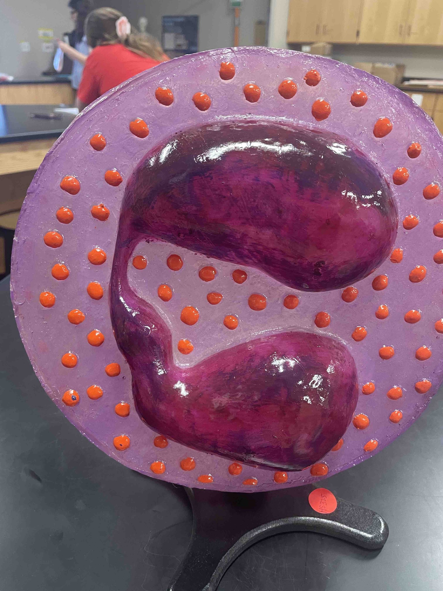
The anatomical model represents an Eosinophil, which is a specialized type of white blood cell (leukocyte) involved in the human immune system. There are two primary morphological features shown in the model that lead to this identification: 1. Bilobed Nucleus: The large, purple structure in the center of the cell is the nucleus. This specific model clearly shows a nucleus divided into two distinct lobes connected by a thin strand of chromatin, which is a classic 'sausage-link' or 'spectacle-shaped' characteristic of eosinophils. 2. Large Cytoplasmic Granules: The orange-red bumps scattered throughout the light purple cytoplasm represent specific granules. In real biological staining (such as Wright's or Giemsa stain), these granules have an affinity for acidic dyes like eosin, which turns them a distinct reddish-orange color. These granules contain basic proteins and enzymes used to fight parasitic infections and mediate allergic reactions. Therefore, the combination of a bilobed nucleus and large, eosinophilic (red/orange) granules confirms that this model is an eosinophil.