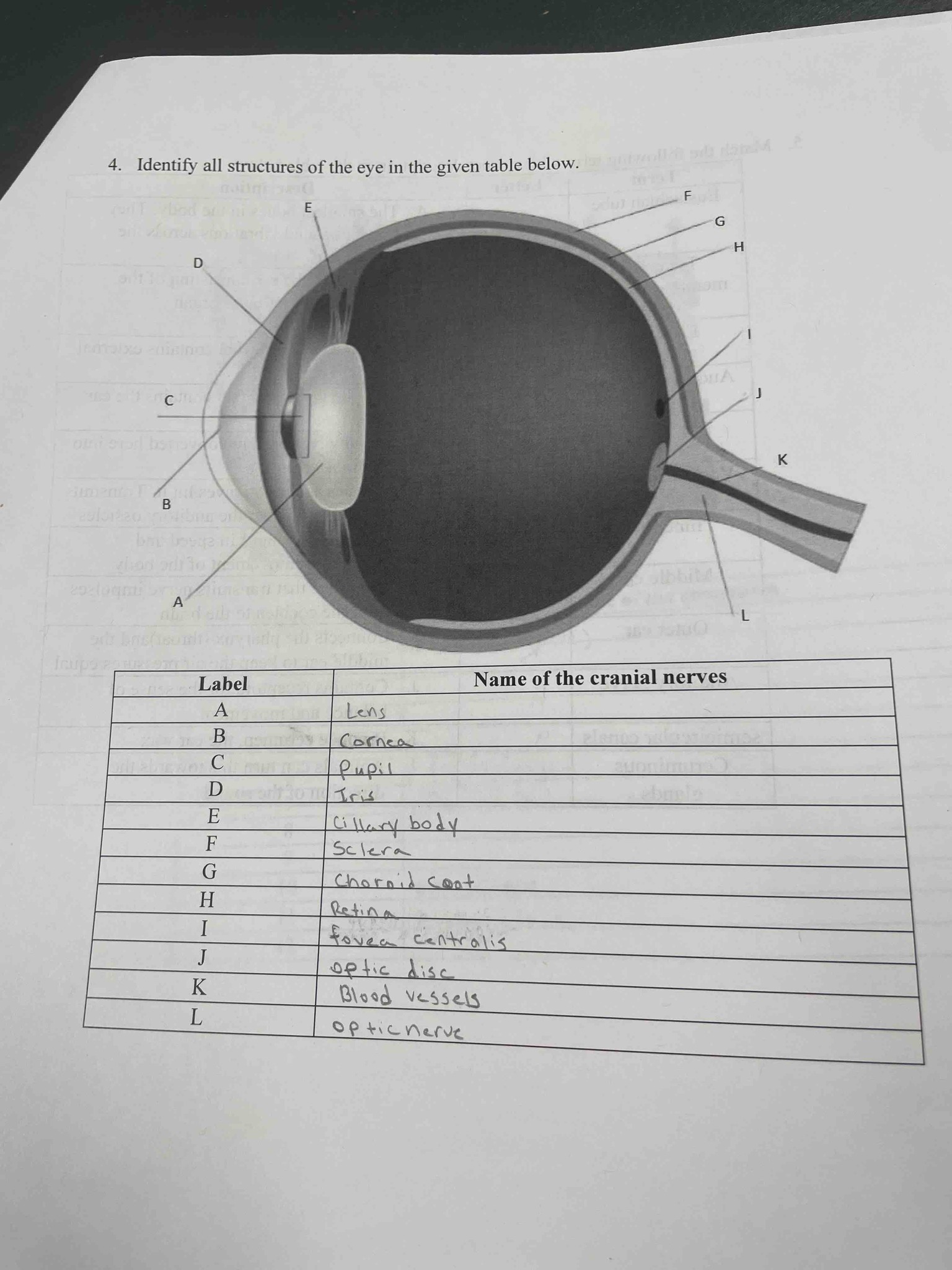
4. Identify all structures of the eye in the given table below. The table requires matching labels A through L to the corresponding anatomical structures of the human eye.
Answer
Based on the anatomical diagram of the human eye, the structures are identified as follows: A is the Lens, the transparent biconvex structure that helps refract light. B is the Cornea, the clear outer layer at the front of the eye. C is the Pupil, the opening in the center of the iris. D is the Iris, the colored part of the eye that controls light entry. E refers to the Ciliary body, which contains muscles that control the shape of the lens. F is the Sclera, the tough white outer layer of the eyeball. G is the Choroid (or Choroid coat), the vascular layer between the retina and the sclera. H is the Retina, the light-sensitive inner lining at the back of the eye. I is the Fovea centralis, a small pit in the retina that provides the clearest vision. J is the Optic disc, also known as the blind spot, where the optic nerve leaves the eye. K represents the Blood vessels (central retinal artery and vein) that supply and drain the eye. L is the Optic nerve (Cranial Nerve II), which transmits visual information from the retina to the brain.