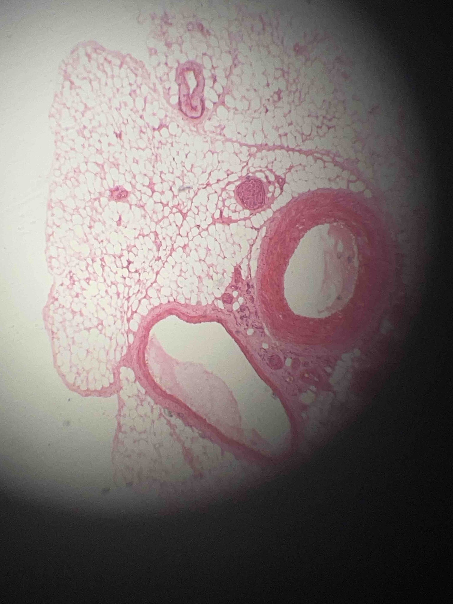
Identify the histological features shown in the micrograph and describe the structures present.
Answer
The micrograph displays a cross-section of neurovascular tissue embedded in adipose tissue (fat). Several key structures can be identified: 1. Adipose Tissue: The majority of the field is filled with large, clear, polygonal cells called adipocytes. They appear clear because the lipid droplet that fills the cell is washed away during the H&E staining process, leaving only a thin rim of cytoplasm and a flattened nucleus at the periphery. 2. Artery: The large circular structure on the right is a muscular artery. It is characterized by a thick tunica media (composed of smooth muscle cells stained pink) and a well-defined lumen. 3. Vein: The larger, more irregularly shaped vessel at the bottom is likely a vein. Veins typically have thinner walls and a larger, more collapsible lumen compared to arteries of similar size. 4. Nerve: To the left of the artery, there is a smaller, rounded eosinophilic structure which represents a small nerve fascicle, wrapped in its protective perineurium. 5. Arterioles/Venules: Smaller vascular structures are also scattered throughout the adipose tissue. Overall, this image illustrates the typical 'neurovascular bundle' often found in connective tissues throughout the body.