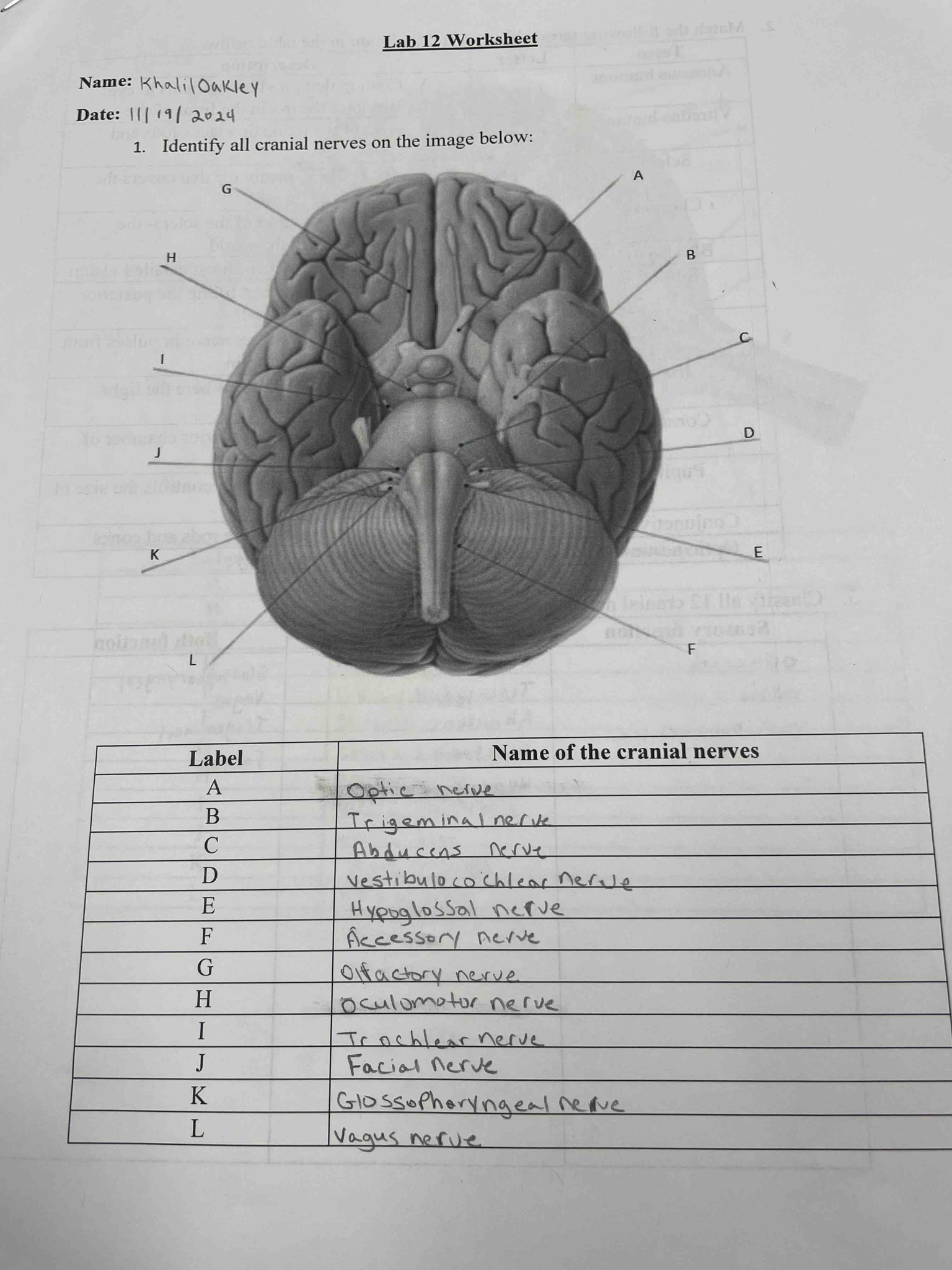
Identify all cranial nerves on the image below:
Answer
Based on the anatomical position on the inferior surface of the brain, the cranial nerves are identified as follows: G is the Olfactory nerve (CN I), located most anteriorly. A is the Optic nerve (CN II), forming the optic chiasm. H is the Oculomotor nerve (CN III), emerging from the midbrain. I is the Trochlear nerve (CN IV), a thin nerve originating posteriorly. B is the Trigeminal nerve (CN V), the large nerve exiting the pons. C is the Abducens nerve (CN VI), emerging from the junction of the pons and medulla. J is the Facial nerve (CN VII) and D is the Vestibulocochlear nerve (CN VIII), both exiting laterally at the cerebellopontine angle. K is the Glossopharyngeal nerve (CN IX), L is the Vagus nerve (CN X), and F is the Accessory nerve (CN XI), which exit sequentially from the lateral medulla. E is the Hypoglossal nerve (CN XII), exiting from the ventral medulla between the pyramid and the olive. The completed table is: A - Optic nerve, B - Trigeminal nerve, C - Abducens nerve, D - Vestibulocochlear nerve, E - Hypoglossal nerve, F - Accessory nerve, G - Olfactory nerve, H - Oculomotor nerve, I - Trochlear nerve, J - Facial nerve, K - Glossopharyngeal nerve, L - Vagus nerve.