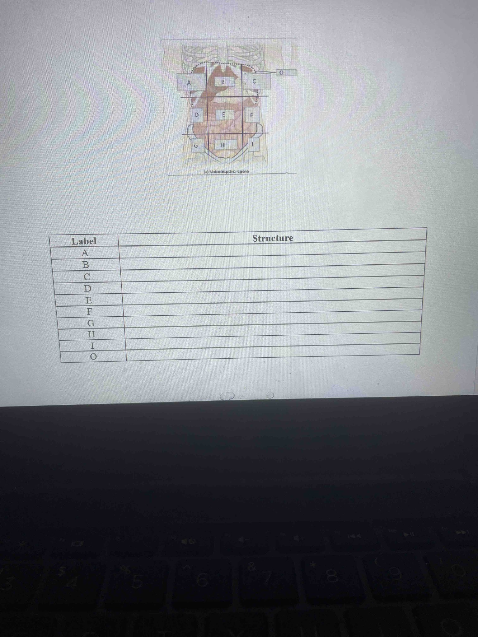
Identify the abdominopelvic regions and the muscle indicated in the provided anatomical diagram. Provide the name for each label from A to I and the label O.
Answer
The image shows the nine abdominopelvic regions of the human body and one primary muscle. The regions are defined by two vertical planes (midclavicular lines) and two horizontal planes (subcostal and transtubercular). Note that anatomical 'right' and 'left' refer to the patient's perspective. The labels are as follows: A = Right hypochondriac region (contains part of the liver and gallbladder); B = Epigastric region (located superior to the stomach); C = Left hypochondriac region (contains part of the stomach and spleen); D = Right lumbar region (contains the ascending colon); E = Umbilical region (surrounds the navel and contains the small intestine); F = Left lumbar region (contains the descending colon); G = Right iliac (inguinal) region (contains the cecum and appendix); H = Hypogastric (pubic) region (contains the urinary bladder); I = Left iliac (inguinal) region (contains the initial part of the sigmoid colon); O = Diaphragm (the dome-shaped skeletal muscle that separates the thoracic cavity from the abdominal cavity).