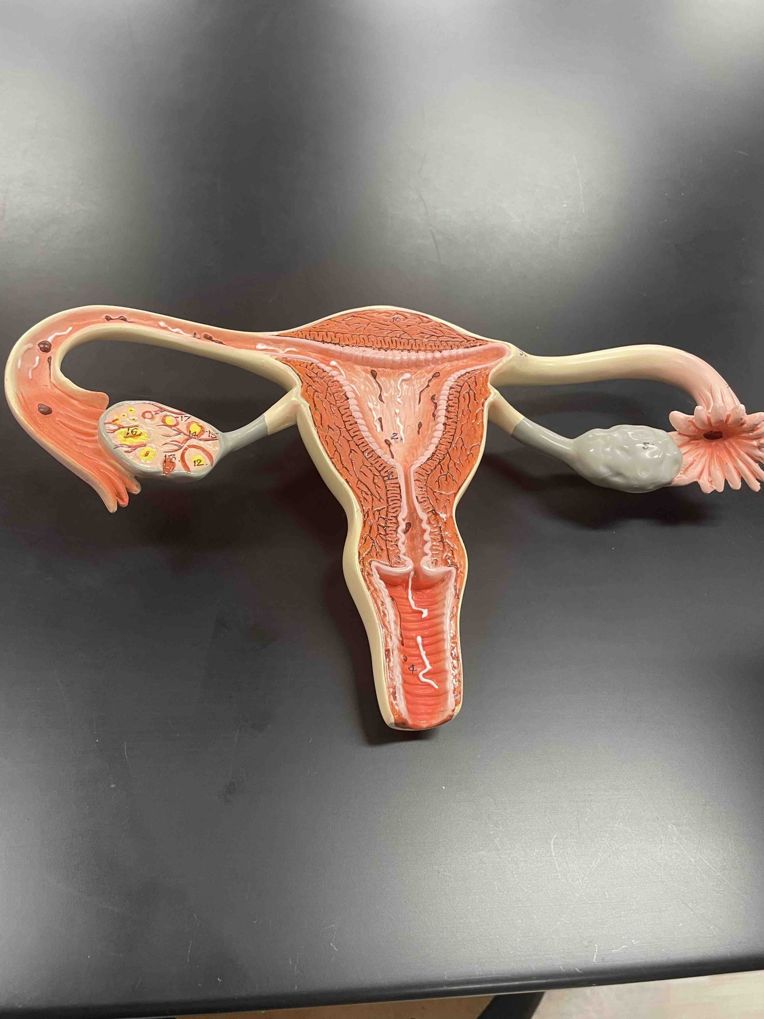
Identify the anatomical parts of the female reproductive system shown in the model, specifically the structures labeled with numbers 4, 12, 16, and the overall organ system.
Answer
The image shows a cross-sectional anatomical model of the human female reproductive system. 1. Overall System: The model displays the uterus, fallopian tubes (oviducts), ovaries, and vagina. 2. Structure 4: This is the vaginal canal (vagina), the muscular tube connecting the external female genitalia to the cervix of the uterus. 3. Structure 12: This points to a corpus luteum within the ovary. This structure is formed from a follicle after an egg has been released and is responsible for producing hormones like progesterone. 4. Structure 16: This identifies an ovarian follicle. Ovaries contain thousands of follicles at different stages of development, each containing an immature egg (oocyte). 5. Other context: The top central portion is the uterus (womb), consisting of the inner endometrium and muscular myometrium. The tubes extending to the sides are the fallopian tubes, which capture the egg from the ovary during ovulation via the fimbriae (the finger-like projections seen on the right).