Identify the anatomical components displayed in the provided medical models and describe their physiological functions in the urinary system.
Answer
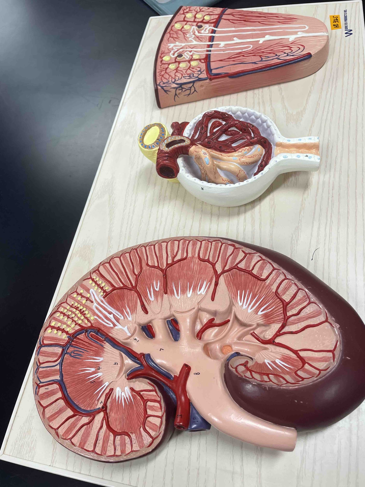
The image displays three different scale models of human kidney anatomy. 1. The large bottom model is a cross-section of the entire kidney. It reveals the outer renal cortex, where ultrafiltration occurs, and the inner renal medulla containing renal pyramids and calyces that collect urine. Major blood vessels like the renal artery (red) and renal vein (blue) are visible, along with the ureter (light pink), which carries urine to the bladder. 2. The middle model is an enlarged view of a renal corpuscle. It shows the glomerulus (a tuft of capillaries) encased within Bowman's capsule (the white cup-like structure). This is the site of blood filtration where water and small solutes are pushed out of the blood into the nephron. 3. The top model is a segment of the renal cortex and medulla showing individual nephrons. This illustrates the complex network of tubules (like the Loop of Henle and distal convoluted tubules) that perform selective reabsorption of nutrients and secretion of waste products, ultimately concentrating the fluid into urine.