Identify the anatomical model and describe the muscles visible in this posterior view.
Answer
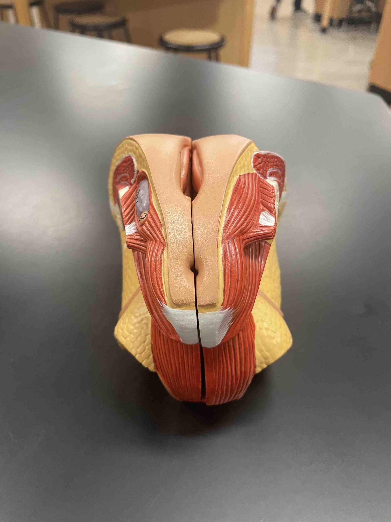
The image shows a posterior (rear) view of an anatomical model of the human larynx (voice box), specifically focusing on the musculature. Key structures visible include: 1. The Arytenoid Muscles: The transverse arytenoid (running horizontally across the back) and the oblique arytenoid (forming an 'X' shape) are visible. These muscles act to adduct the vocal folds, closing the glottis for phonation. 2. Posterior Cricoarytenoid Muscles: These are the large, fan-shaped muscles located on the posterior surface of the cricoid cartilage. They are the only muscles responsible for abducting (opening) the vocal folds. 3. Epiglottis: Seen at the very top of the model. 4. Thyroid Cartilage: The large lateral plates that form the outer 'shield' of the larynx. This view is essential for understanding how the intrinsic muscles of the larynx tilt and rotate the cartilages to control the tension, length, and position of the vocal cords.